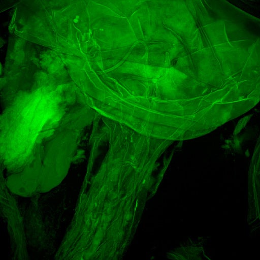
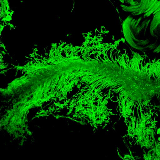
We investigate comparative morphology of Heteroptera using SEM, confocal laser microscopic (CLM), and macroimaging techniques. The image on the left shows part of the female genitalia, the median oviduct and anterior portion of the bursa copulatrix, of Zelus renardii (Reduviidae), as seen in the confocal microscope (CEPCEB at UCR, Leica SP2). The paired tubes arising from the bursa are the ducts of the paired pseudospermathecae. The image on the right documents the glandular portion of the vermiform gland in Triatoma protracta, one of the local species of kissing bugs.
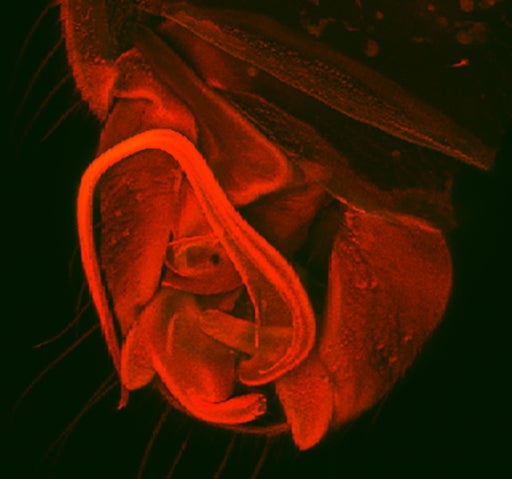
CLM is also used in the lab to document the complex male genitalic features of the tiny (usually 1-3mm body length) Dipsocoromorpha, the litter bugs (see image in the middle). Genitalia in these bugs range from relatively simple with novel symmetrical abdominal appendages (some Ceratocombidae), to strong sinistral (some Dipsocoridae) or dextral (Schizopteridae) asymmetries that frequently include tremendous asymmetrical modifications of the remaining abdominal segments. The image shows male genitalic structures of one species of Schizopteridae: Schizopterinae, as seen in the CLM.


